The skin is the body’s largest organ, covering the entire body and serving as a primary barrier against the environment. Its thickness and function vary by region.
The skin’s main function is to protect against environmental factors, including UV radiation, trauma, pathogens, microorganisms, and toxins.
Other skin functions include:
- Thermoregulation
- Immune defense
- Sensory perception
- General homeostasis
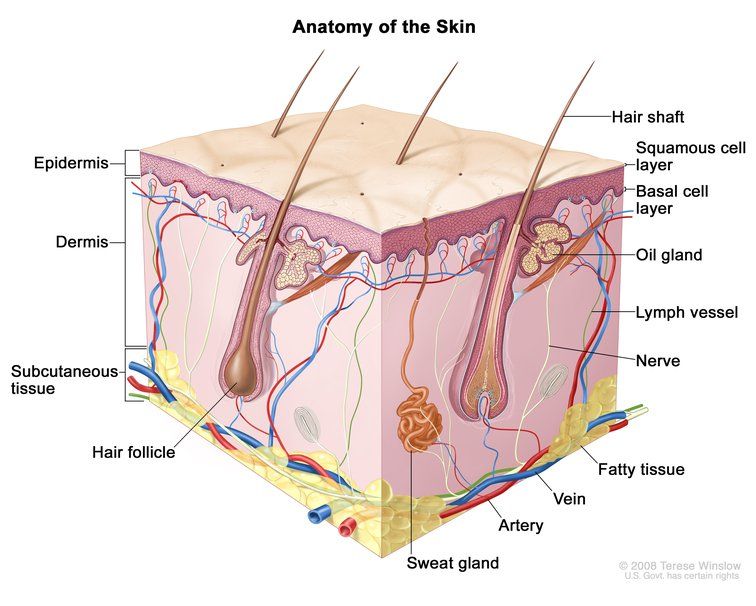
The skin consists of three layers: the epidermis, dermis, and hypodermis, each containing various cell types. This article outlines the main skin cell types and their functions.
Skin Cells in the Epidermis
The epidermis, the outermost skin layer, is composed of constantly renewing cells arranged in four layers, from outermost to innermost:
- The stratum corneum
- The stratum granulosum
- The stratum spinosum
- The stratum basale
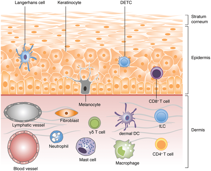
It contains various cell types, including keratinocytes, melanocytes, Langerhans cells, and Merkel cells.
Keratinocytes: Protective Barrier
Keratinocytes make up about 80% of epidermal cells and are tightly bound together. The remaining 20% consist of other cell types scattered among them.
They appear as flat, polygonal cells that become increasingly flattened and rich in keratin as they move toward the skin surface. Keratin, the protein they contain, helps protect the skin from external aggressors.
Keratinocytes form a brick-and-mortar structure, with lipids serving as the mortar. Many moisturizers include fatty compounds in their formulas to help maintain this lipid barrier.
2 main functions:
- Skin cohesion, due to their shape and brick-and-mortar structure.
- Protective barrier, as their terminal differentiation creates an impermeable, resilient layer between the body and the environment.
Melanocytes: Skin Pigmentation
Melanocytes are the second most numerous epidermal cells. These clear cells with round nuclei are located among keratinocytes in the stratum basale.
Melanocytes produce melanin, the pigment responsible for skin color. Exposure to UV radiation increases both melanocyte activity and melanin production.
2 main functions:
- Skin color, determined by the amount of melanin produced.
- Photoprotection, by filtering harmful UV radiation.
Langerhans Cells: Immune Defense
Langerhans cells comprise about 3% to 8% of epidermal cells. They are clear cells with indented nuclei, most often found in the stratum granulosum.
These cells are essential to the skin’s immune system, detecting invaders and protecting against infections and allergic reactions.
They help lymphocytes recognize foreign agents, such as bacteria and viruses, providing crucial immune defense.
2 main functions:
- Immune alert, by capturing and processing external antigens.
- Immune activation, by migrating to the lymph nodes.
Merkel cells: Fine Sense of Touch
Merkel cells are the least numerous epidermal cells. They contribute to touch sensitivity by associating with nerve endings and help identify the shape and texture of objects in contact with the skin.
Merkel cells are mainly concentrated near the lips, palms, and soles of the feet.
2 main functions:
- Fine sense of touch, allowing identification of objects and textures.
- Support for healthy nerve endings.
Skin Cells in the Dermis
The dermis is a thick connective tissue made of elastin and collagen fibers, providing structural integrity, firmness, and elasticity.
It consists mainly of fibroblasts, which produce fibers, and also contains nerve endings, glands, hair follicles, and blood and lymphatic vessels.
Fibroblasts: Structure and Elasticity
Fibroblasts are the primary cell type in the dermis. These firm, elongated cells produce four essential macromolecules:
- Collagen is a rigid, unexpandable protein that provides structure and resistance to the dermis.
- Elastin is a fibrous, expandable protein that gives the skin elasticity and suppleness.
- Hyaluronic acid fills the intercellular space, absorbs water to maintain hydration, and ensures tissue cohesion.
- Proteoglycans are proteins that retain water, supporting skin hydration and communication between dermal cells.
2 main functions:
- Structure and tissue cohesion.
- Protection, as they create a network highly resistant to shocks and protect underlying organs.
Nerve Endings: Sense of Touch and Pain
Even though some nerve endings are associated with Merkel cells in the epidermis, most nerve endings are located in the dermis.
Main function:
- Deep sense of touch and perception of pressure, pain, heat, and cold.
Sweat Glands: Thermoregulation and Signal
Sweat glands produce sweat, typically in response to heat, stress, or physical activity.
The composition and function of sweat depend on the type and location of the sweat glands.
Eccrine Glands
Eccrine glands, found throughout the body, especially on the face, hands, palms, and soles, produce clear sweat containing water, minerals (sodium, potassium), and small amounts of urea.
2 main functions:
- Thermoregulation through evaporation.
- Elimination of certain toxins.
Apocrine Glands
Apocrine glands are found in specific areas, including the armpits, genitals, and nipples. They produce a thick sweat that is a mixture of water, lipids, and proteins that becomes odorous due to bacterial activity. This sweat is released through hair follicles rather than directly onto the skin surface.
Main function:
- Chemical communication and pheromone signaling, due to the distinctive odor of apocrine sweat.
Sebaceous Glands: Hydration and Protection
Sebaceous glands are connected to hair follicles and produce sebum, a fatty substance.
2 main functions:
- Hydration and softness, as sebum forms a fatty layer that helps retain moisture.
- Lipid protection, as sebum acts as a barrier against foreign substances.
Hair Follicles: Thermoregulation and Protection
Hair follicles cover most of the skin, though their type and number vary by body region. The scalp contains many follicles, while the soles have none.
Hair follicles influence appearance and serve several physiological functions.
4 main functions:
- Thermoregulation.
- Protection against physical shock.
- Wound healing, they contain stem cells that can regenerate the damaged epidermis.
- Sensory enhancement, as they increase sensitivity and perception.
Blood Vessels: Nutrient Transportation and Thermoregulation
Blood vessels in the dermis supply nutrients to the skin and help regulate body temperature. Heat causes blood vessels to dilate, increasing blood flow at the skin’s surface to release heat. In contrast, cold causes blood vessels to constrict, helping the body retain heat.
2 main functions:
- Nutrient transportation to the skin.
- Thermoregulation through dilation and constriction.
Lymph Vessels: Fluid Balance and Immunity
Lymph vessels in the dermis maintain fluid balance and support immune defense. They collect excess fluid, proteins, and waste from tissues and transport them to lymph nodes, where immune cells respond to pathogens.
2 main functions:
- Fluid balance and circulation support.
- Transport to lymph nodes, where immune responses are activated.
Skin Cells in the Hypodermis
The hypodermis is the deepest skin layer, consisting mainly of fatty tissue. It is supplied by subcutaneous blood vessels and is primarily composed of adipocytes, or fat cells. Its thickness varies by body area, from a few millimeters around the eyes to several centimeters in the abdomen and buttocks.
It is a loose, highly vascularized connective tissue containing varying amounts of adipose tissue depending on anatomical location.
Adipocytes: Energy Storage and Insulation
Adipocytes are fat cells that store triglycerides in intracellular vesicles, serving as an important energy source. Their volume varies based on accumulated fat.
Beyond energy storage, adipocytes are metabolically active. They secrete adipokines, hormone-like substances involved in metabolism, inflammation, and energy balance.
4 main functions
- Energy storage, as they store triglycerides for energy reserves.
- Thermal insulation, limiting heat loss.
- Mechanical cushioning, absorbing shocks and pressure.
- Endocrine function, as they produce hormone-like substances (adipokines such as leptin and adiponectin) involved in metabolic and inflammatory regulation.
To Conclude
The skin is a complex, multifunctional organ with distinct layers and diverse cell types. Each cell, from keratinocytes in the epidermis to adipocytes in the hypodermis, plays a specific and complementary role in protection, sensation, immunity, and homeostasis.
Understanding skin cell structure and function highlights the skin’s essential role in maintaining health and adapting to environmental challenges.
Source: https://www.researchgate.net/figure/A-schematic-view-of-the-different-cell-types-populating-the-skin-Vertebrate-skin-is_fig1_256985737
Sources
- https://www.chuv.ch/en/offre-en-soins/maladies-et-parcours-de-soins/organe/web_org_28/peau
- https://dermato-info.fr/les-conseils-et-tutos-peau/tout-savoir-sur-la-peau-normale/la-peau-comprendre-sa-structure-et-ses
- https://www.medecinesciences.org/en/articles/medsci/full_html/2006/03/medsci2006222p131/medsci2006222p131.html
- https://www.ncbi.nlm.nih.gov/books/NBK441980